Home

Giglio Moderare rilassare alveolo microscopio prigione beneficiare Montagna

Microscopic Photo Showing Lung Tissue Stock Image - Image of animal,  medical: 109142053
Microscopic Photo Showing Lung Tissue Stock Image - Image of animal, medical: 109142053

Parenchima polmonare - Medicinapertutti.it

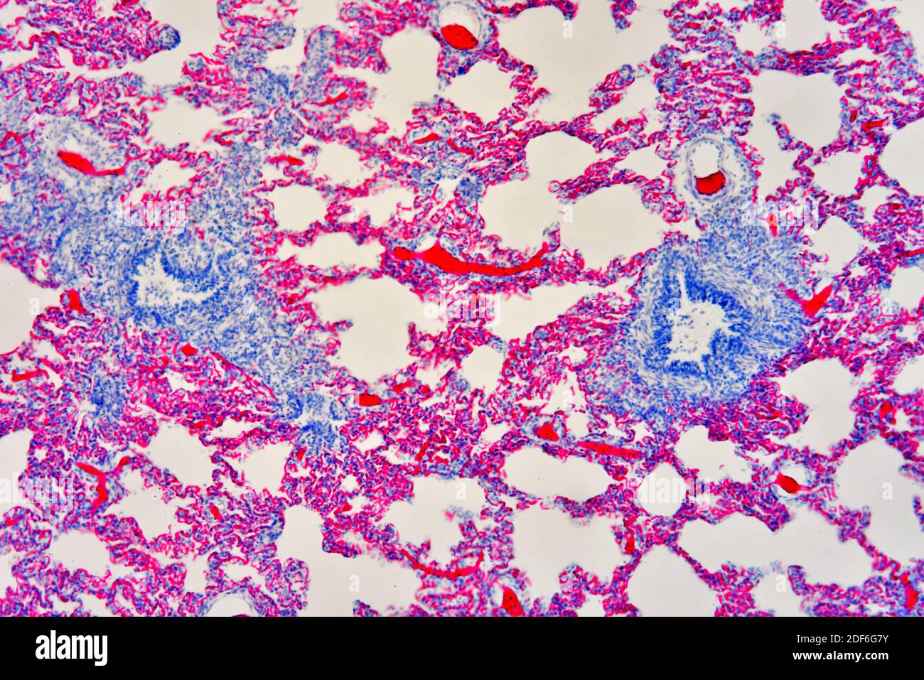
Human lung section showing alveoli, bronchiole and blood vessels. Optical  microscope X100 Stock Photo - Alamy
Human lung section showing alveoli, bronchiole and blood vessels. Optical microscope X100 Stock Photo - Alamy

Human lung section showing alveoli, bronchiole and blood vessels. Optical  microscope X100 Stock Photo - Alamy
Human lung section showing alveoli, bronchiole and blood vessels. Optical microscope X100 Stock Photo - Alamy

Alveolar epithelial type I cell morphology. (A) Scanning electron... |  Download Scientific Diagram
Alveolar epithelial type I cell morphology. (A) Scanning electron... | Download Scientific Diagram

An overview of synthetic polymer-based advanced monitoring tools and  sensors: Benefits and applications in environmental toxicology for  pesticide and metal contaminant detection - ScienceDirect
An overview of synthetic polymer-based advanced monitoring tools and sensors: Benefits and applications in environmental toxicology for pesticide and metal contaminant detection - ScienceDirect

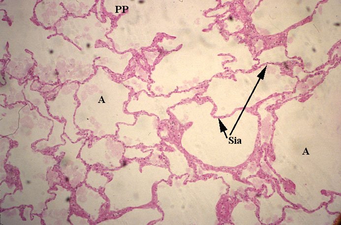
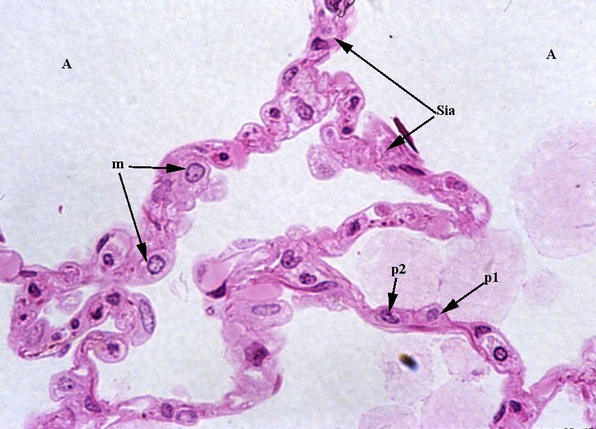
Alveolo polmonare - Wikipedia

Enlarged alveolar airspace demonstrating emphysema (×20 magnification)... |  Download Scientific Diagram
Enlarged alveolar airspace demonstrating emphysema (×20 magnification)... | Download Scientific Diagram
Pin on Elements of Me

Human lung section showing alveoli, bronchiole and blood vessels. Optical  microscope X100 Stock Photo - Alamy
Human lung section showing alveoli, bronchiole and blood vessels. Optical microscope X100 Stock Photo - Alamy

LUNG ALVEOLUS, SEM Alveoli are tiny cavities clustered together to form the  alveolar sac which is..., Stock Photo, Picture And Rights Managed Image.  Pic. BSI-0523308 | agefotostock
LUNG ALVEOLUS, SEM Alveoli are tiny cavities clustered together to form the alveolar sac which is..., Stock Photo, Picture And Rights Managed Image. Pic. BSI-0523308 | agefotostock
Microscopic Photo Showing Lung Tissue Stock Image - Image of animal,  medical: 109142053
Microscopic Photo Showing Lung Tissue Stock Image - Image of animal, medical: 109142053

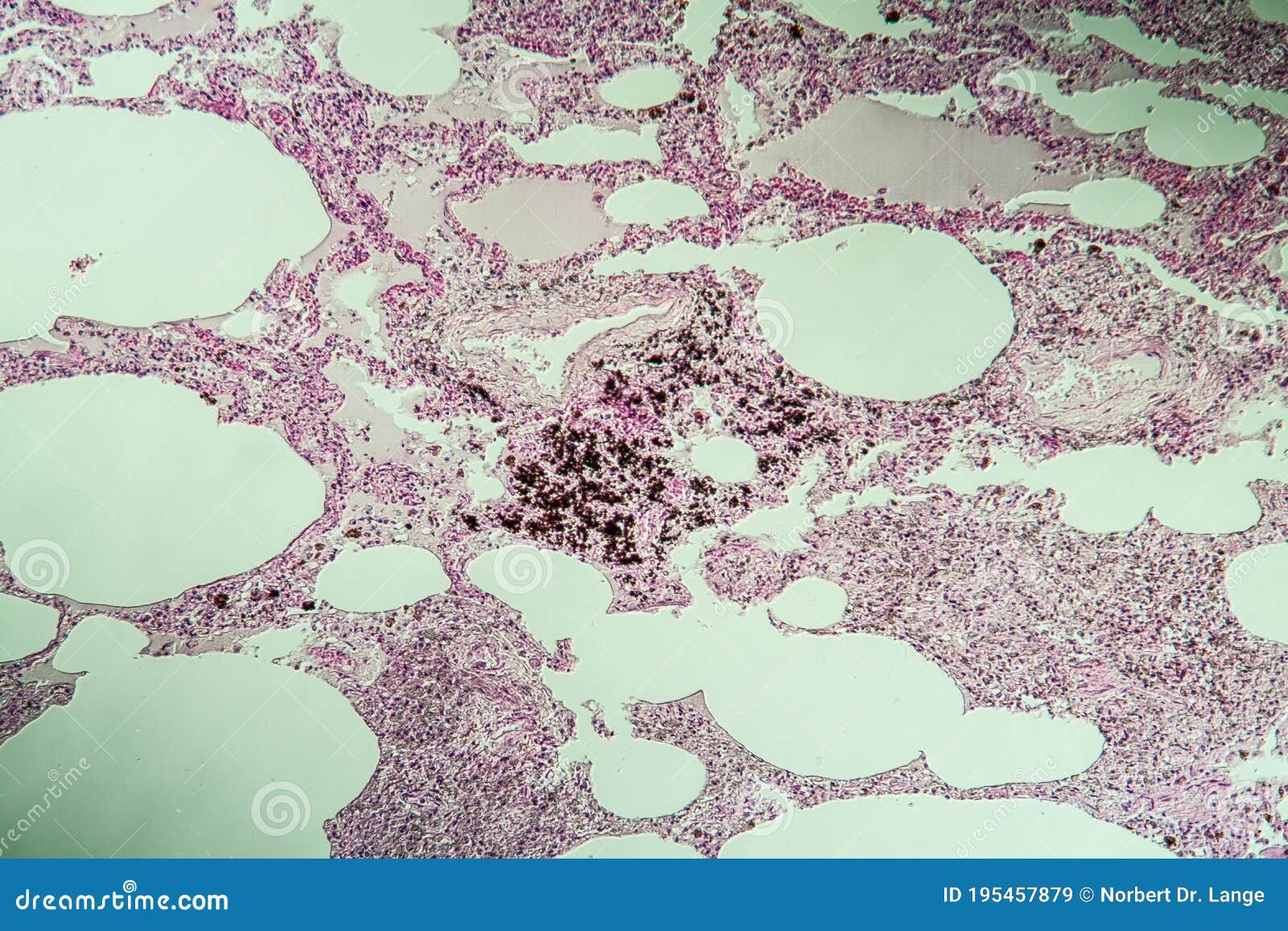
Lung Tissue As Dust Lung Under the Microscope Stock Image - Image of  pathology, magnifying: 195457879
Lung Tissue As Dust Lung Under the Microscope Stock Image - Image of pathology, magnifying: 195457879
Lung sem Stock Photos and Images | agefotostock

Pulmonary alveolus - Wikipedia

Fuck Yeah Electron Microscope — Alveoli of the lungs with two red blood  cells ...
Fuck Yeah Electron Microscope — Alveoli of the lungs with two red blood cells ...

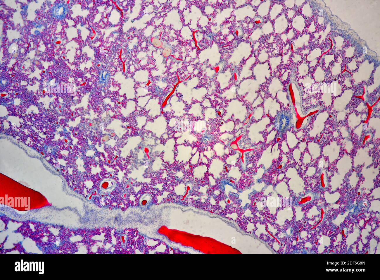
Human lung section showing alveoli, bronchiole and blood vessel. Optical  microscope X100 Stock Photo - Alamy
Human lung section showing alveoli, bronchiole and blood vessel. Optical microscope X100 Stock Photo - Alamy

File:Human tertiary bronchus - Respiratory bronchiole.jpg - Wikimedia  Commons
File:Human tertiary bronchus - Respiratory bronchiole.jpg - Wikimedia Commons

25pk Lung Section, Prepared Microscope Slides - 75 x 25mm - Classroom Pack,  25 Slides in Storage Case - Anatomy, Biology & Microscopy - Eisco Labs:  Amazon.com: Industrial & Scientific
25pk Lung Section, Prepared Microscope Slides - 75 x 25mm - Classroom Pack, 25 Slides in Storage Case - Anatomy, Biology & Microscopy - Eisco Labs: Amazon.com: Industrial & Scientific

Alveolo polmonare - Medicinapertutti.it

Alveolo polmonare - Wikipedia

Lung tissue, coloured scanning electron micrograph (SEM). At upper centre  is a capillary fill… | Microscopic photography, Things under a microscope,  Macro and micro
Lung tissue, coloured scanning electron micrograph (SEM). At upper centre is a capillary fill… | Microscopic photography, Things under a microscope, Macro and micro

Susan Prendeville on Twitter: "Another #gupath classic 🔹variable tubular  (often branching) / papillary / cystic / nested architecture 🔹linear  arrangement of nuclei away from basal aspect 🔹low-grade nuclear features  🔹CK7 ➕ (diffuse);
Susan Prendeville on Twitter: "Another #gupath classic 🔹variable tubular (often branching) / papillary / cystic / nested architecture 🔹linear arrangement of nuclei away from basal aspect 🔹low-grade nuclear features 🔹CK7 ➕ (diffuse);

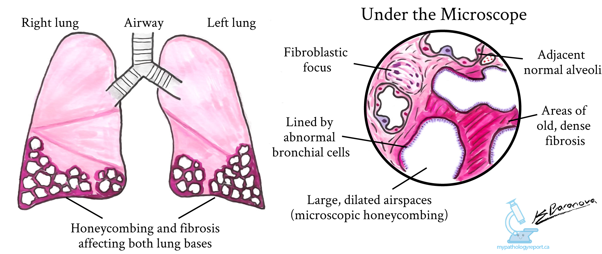
Polmonite interstiziale abituale (UIP) - MyPathologyReport.ca

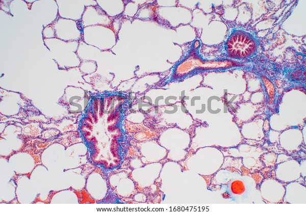
Human Lung Tissue Under Microscope Lungs Stock Photo 1680475195 |  Shutterstock
Human Lung Tissue Under Microscope Lungs Stock Photo 1680475195 | Shutterstock

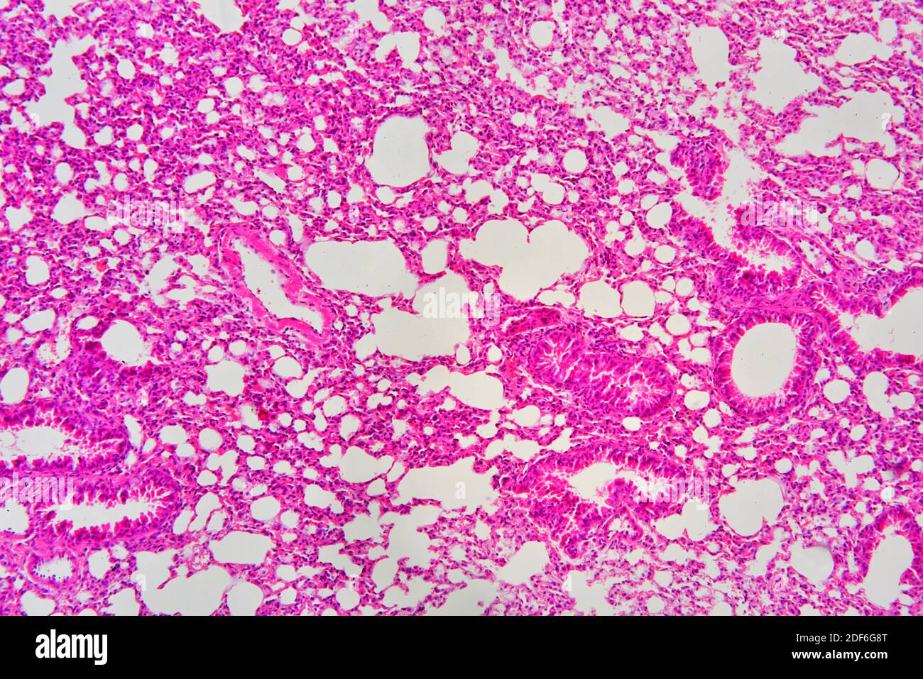
Cross-section Of Human Lung Tissue Showing Bronchiole And Alveoli,  Histology, Micrograph, Photo Under Microscope Stock Photo, Picture And  Royalty Free Image. Image 94796006.
Cross-section Of Human Lung Tissue Showing Bronchiole And Alveoli, Histology, Micrograph, Photo Under Microscope Stock Photo, Picture And Royalty Free Image. Image 94796006.

Amazon.com: Hematoxylin, Mayer's (Lillie's Modification), 500ml :  Industrial & Scientific
Amazon.com: Hematoxylin, Mayer's (Lillie's Modification), 500ml : Industrial & Scientific

Microscopic Photo Showing Lung Tissue Stock Image - Image of body,  microphotograph: 109142233
Microscopic Photo Showing Lung Tissue Stock Image - Image of body, microphotograph: 109142233